Also Known As
Thrombophilia
This article was last reviewed on
This article waslast modified on 29 September 2023.
What is it?

A hypercoagulable disorder, also known as thrombophilia, is an inherited or acquired condition that increases the risk of developing inappropriate or excessive thrombus (blood clot) formation. In general within the population acquired hypercoagulable disorders are more common than inherited disorders.

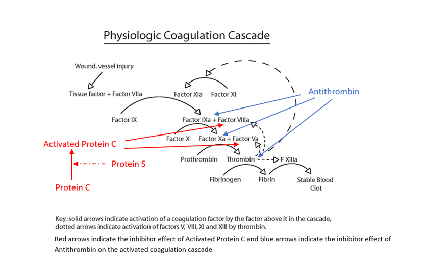
Blood clotting is normal response to blood vessel or tissue injury. Blood is normally in a fluid state (anticoagulated) within the body to enable flowing through a vessel network of arteries, capillaries and veins, delivering oxygen and nutrients to the organs of the body and removing carbon dioxide and waste products. When a blood vessel is injured, it begins to leak blood, either externally e.g a skin cut or internally into body tissues e.g a bruise. The body stops this blood loss through a complex clotting process called haemostasis. During haemostasis, the injured blood vessel becomes narrower (constricts) to reduce blood flow, cells in the blood called platelets stick (adhere) to the injury site and clump together to form a loose platelet plug and the coagulation cascade is triggered (initiated). During the cascade process, the body sequentially activates coagulation factors, which are proteins that create a net of fibrin threads, which weave them through the platelet plug making a stable firm fibrin blood clot, whilst still allowing blood to flow through the damage vessel. This blood clot covers the injured area and forms a barrier to prevent further blood loss and the clot should stay in place until the injury has healed.

In normal haemostasis once the body has activated the clotting process, there are regulatory feedback mechanisms, which limit and control the clotting process (natural anticoagulants), prevent the complete blocking of the vessel by excessive clot formation and by removal of the fibrin clot once the injury has healed (fibrinolysis). Hypercoagulable disorders can occur when something goes wrong within this clotting process. If the clotting process activates inappropriately, or feedback mechanisms fail to work effectively to limit formation or removal of fibrin clot, then there can be inappropriate and/or excessive blood clot formation.

Presentation of Hypercoagulable Disorders 

Thrombosis can occur within veins or arteries, however the mechanism of clot formation is different, with venous thrombosis associated with sluggish movement of blood (stasis) or imbalance of the clotting progress and feedback mechanism, whereas arterial thrombosis more commonly results from the rupture of an atherosclerotic plaque due to build-up of cholesterol in the arterial wall. Blood clots are referred to as thrombi (one = thrombus) when they form in a blood vessel; thrombi may break off and block another blood vessel in another part of the body, where they are referred to as emboli (one =embolus).

  • Venous thromboembolism (VTE) is the most common condition associated with hypercoagulable disorders, with blood clots most frequently forming in the deep veins of the legs (DVT) causing redness, pain and swelling particularly at the back of legs. However, a DVT can become life-threatening if the clot breaks free and travels to other parts of the body through the bloodstream, particularly if it becomes lodged in the arteries of lung, called a pulmonary embolism PE or to the brain causing strokes.
  • Although less common, thrombosis can occur in unusual venous circulation sites causing cerebral vein thrombosis (brain), hepatic and portal vein thrombosis (liver), mesenteric vein thrombosis (small intestines), renal vein thrombosis (kidney), venous thrombosis in arms and ovarian vein thrombosis
  • Recurrent foetal loss (miscarriages) and other complications in pregnancy may be associated with thrombophilia.
  • Pupura fulminas is the result of severe (homozygous) Protein C or Protein S deficiency causing massive thromboembolic complications (a form disseminated intravascular coagulation) in newborns shortly after birth. It is life-threatening causing tissue death (necrosis) and bleeding under the skin and other organs, without treatment with Protein C concentrate or fresh frozen plasma. It can also occur in adults associated with acquired rapid onset following acute infections.
  • Warfarin medication may also produce a similar phenomenon to purpura fulminas during the early days of starting therapy due to reducing the level of Protein C which has a short half-life, before significant falls in the other vitamin K dependent procoagulants occurs.
  • Disseminated intravascular coagulation (DIC) is a life-threatening, acute, acquired condition that causes tiny clots throughout the body,often associated with sepsis. It uses up coagulation factors and platelets at an accelerated rate, leading to both formation of thrombi in many organs and bleeding.
  • Immunothrombosis is one of the significant complications of severe COVID-19 infections is a coagulopathy that seems to be related to the occurrence of venous and arterial thromboembolic disease, with strong local pulmonary thrombotic microangiopathy and direct endothelial cell infection and injury by the virus. The coagulation changes mimic but are not identical and do not meet the criteria for disseminated intravascular coagulation.

Risk factors for Hypercoagulable Disorders 

Most people have the potential to develop a blood clot but there are several situations or conditions that can increase an individual’s risk of a hypercoagulable disorder including:

  • Inherited condition that increases the risk of thrombosis or family history of thrombosis
  • Decreased blood flow due to prolonged periods of immobility
  • Admission to hospital
  • Injury or surgery affecting a vein
  • Increased oestrogen due to pregnancy or hormone therapy
  • Medical conditions linked with cancer, inflammatory disorders, heart failure, atrial fibrillation, hypertension, antiphospholipid syndrome and obesity
  • Increasing age

Certain inherited gene mutations that may predispose someone to hypercoagulable states, such as factor V Leiden or the prothrombin G20210 A mutation, are relatively common in the population, but it is thought that they add only a slight increase in the risk of actually developing a problem with clotting. Other inherited hypercoagulable disorders, such as protein C deficiency, protein S deficiency, and antithrombin deficiency have a higher risk of thrombosis but are relatively rare. These are generally due to genetic mutations that lead to a deficiency or dysfunction in the coagulation protein that the gene produces. All of the inherited disorders (except for antithrombin deficiency) may be seen in heterozygous (one gene copy) or homozygous (two gene copies) form. If someone has two mutated gene copies, they tend to have a more severe form of the condition, and if they are heterozygous in more than one condition, the risk of clotting tends to be additive (and sometimes they multiply the risk). With inherited hypercoagulable disorders, the first thrombotic episode may be seen at a relatively young age (less than 40 years of age). 

Accordion Title
About Hypercoagulable Disorders
  • Activation Problems

    Hypercoagulable Disorders effect on clotting process

    Increased Activation Problems

    Problems with coagulation activation

    Anything that interrupts the smooth blood vessel walls, essential for constant blood flow, may increase the risk of activating the coagulation cascade.

    • Blood vessel wall abnormalities (usually combined with a clotting abnormality, possibly associated with von Willebrand factor)
    • Atherosclerosis -- the build-up of cholesterol, lipid and calcium deposits in the walls of arteries. They make the blood vessel walls less smooth, weaken them, and eventually form plaques that may rupture and lead to abnormal platelet activation and clots associated with strokes and heart attacks.
    • Vasculitis -- inflammation of blood vessel walls may increase the risk of platelet adhesion and coagulation factor activation. Healed vasculitis may provide sites within the vessels for accelerated atherosclerosis.
    • Regulation/feedback mechanism problems


  • Regulation problems

    Problems with coagulation cascade regulation

    Inherited:

    • Factor V Leiden is a condition caused by mutation in Factor V(F5) gene at position 1691, increasing the risk of Deep Vein Thrombosis (DVT) and Pulmonary Embolism (PE) by making activated Factor V resistant to the anticoagulant effect of Activated Protein C, which regulates the clotting process by inhibiting activated factors Va and VIIIa Studies show that around 3-8% of Caucasians have one copy of mutated FV gene (heterozygous) and 1 in 5000 Europeans have 2 copies of this gene mutation (homozygous). Factor V Leiden mutation is rarer in black Africans, Asians, Australian and American natives.
    • Prothrombin 20210 mutation is caused by mutation in prothrombin gene (factor II) at position 20210 in 3’ untranslated gene region. This increases the circulating level of Factor II (thrombin) which is a major factor in the clotting cascade at promoting clotting increasing the blood tendency to form blood clots. Recently, this mutation has also been associated with reduced reactivity to antithrombin, leading to antithrombin resistance. . This mutation is prevalent in Caucasians.
    • Plasminogen deficiency is a rare congenital disorder. Plasminogen is activated to form plasmin. Plasmin helps break apart the clot's cross-linked fibrin net. However, there are so few cases of deficiency, that any association with VTE is unclear and published literature is conflicting on the thrombotic risk associated with Plasminogen deficiency.

    Inherited (rare) or Acquired (see diagram below)

    • Antithrombin deficiency or dysfunction - (normal level of a structurally abnormal molecule). - Antithrombin is the most important natural anticoagulants, reducing (inhibiting) the activity of activated clotting factors Xa, IXa, XIa, and thrombin. Deficiency can lead to a clot formation.
    • Protein C, and/or co-factor Protein S deficiency or dysfunction. The role of the natural anticoagulants with the Protein C pathway is to generate Activated Protein C which reduces (inhibits) the activity of factor VIIIa and factor Va. Deficiency can lead to increased clot formation and more serious complications as previously described.
    • Activated Protein C resistance (APCR), there are several other rarer FV genetic mutations that can also cause APCR. Also high levels of factor VIII and presence of a lupus anticoagulant can result in an acquired defect.
    Physiologic Coagulation Cascade


    • Antiphospholipid syndrome (lupus anticoagulant and anticardiolipin antibody) is an acquired condition caused by antibodies developed against phospholipids which are molecules that participate in the coagulation cascade. These antibodies include lupus anticoagulants (LA), anticardiolipin antibodies and antibodies to beta 2 gycoprotein-1. This is considered as an autoimmune condition causing arterial or venous thrombosis, miscarriages in first trimester of pregnancies and may be associated with systemic lupus erythematosus.

    Problems involving platelets

    Acquired:

    • Heparin-induced Thrombocytopenia (HIT) is a condition where a patient receiving an anticoagulant called heparin produces antibodies against the heparin which bind to platelet factor 4 forming a heparin/PF4 complex whichactivate platelets and produce a hypercoagulable state. causing venous or arterial thrombosis. This condition is most commonly association with the use of unfractionated heparin which is used less frequently now in clinical practice.
    • Vaccine induced immune thrombocytopenia and thrombosis (VITT) is rare but is caused by anti-platelet factor 4 (PF4) antibodies that arise following covid-19 vaccination and lead to intense activation of platelets and the coagulation system. Thrombosis affects the cerebral veins in 50% of cases, but any arterial or venous vascular bed may be involved and around one third of patients have thrombosis in multiple sites. Thrombotic Thrombocyopenia Purpura (TTP) usually acquired but can rarely be inherited. Regulation and breakdown of von Willebrands factor (VWF) from a large protein to smaller subunits (multimers) is by ADAMTS-13. Autoantibodies formed to ADAMTS-13 result in excessive large VWF multimers in the circulation which are hyper-adhesive and actively bind to platelets, producing thrombi.
    • Disseminated intravascular coagulation (DIC) – see previous
    • Immunothrombosis – see previous
  • Breakdown problems

    Problems with clot breakdown (fibrinolysis)

    Inherited and/or Acquired:

    • Plasminogen deficiency – see above
    • Dysfibrinogenaemia -can be an inherited or acquired condition which results in an abnormal fibrinogen molecule which results in interference with forming a normally structured fibrin clot and/or clot resistance to lysis by plasmin. This can lead to both bleeding and thrombotic complications.
  • Other factors

    Other factors associated with an increased risk of inappropriate blood clot formation:

    • Cancer – Cancers may cause hypercoagulable states for a variety of reasons.: 1 Tumour growth may cause external compression on a blood vessel, or in some cases, actually extend into the vasculature (for example, renal cell cancers extending into the renal veins). 2 Cancers are often associated with inflammation and immobility. 3 Some cancers release substances which initiate clotting. 4). Treatments for cancer (radiation, chemotherapy) may leave patients more susceptible to hypercoagulation.
    • Paroxysomal Nocturnal Haemoglobinuria (PNH) results from a mutation in a gene called PIGA. PIGA gene products normally protect red blood cells from complement system. The mutation causes increased thrombosis and haemolytic anaemia due to overactivation of the complement system on the surface of red blood cells.
    • Myeloproliferative neoplasms (formerly known as myeloproliferative disorders) result in excess blood cell production, resulting in polycythaemia vera (increased red blood cell production) and essential thrombocythaemia (increased platelet production). This results in an increased risk of thrombosis.
    • Venous stasis describes any situation that immobilizes someone for long periods of time, such as prolonged bed rest with an illness or after surgery. Immobility may lead to slow or restricted blood flow (venous stasis) and an increased risk of developing a blood clot especially in the deep veins of the legs (DVT). This can also occur in people travelling on long journeys while immobilised in aeroplanes and coaches
    • Recent surgery with trauma to tissue and blood vessels
    • Atrial fibrillation (rapid, uneven heartbeat) – associated with an increased risk of stroke
    • Heart Failure - may cause slowing of blood flow (stasis)
    • Obesity
    • Pregnancy, which naturally causes a hypercoagulable state, combined oral contraceptives (“the pill”) and hormone replacement therapy
    • Elevated levels of homocysteine which can be an inherited defect but also acquired linked with folate deficiency can increase thrombotic risk, how is uncertain but may be relate to interference with vessel endothelial cell function. It has been associated with a slightly increase the risk of a heart attack.
    • High levels of certain clotting factors including of FVII, FVIII, FIX,FXI, and fibrinogen may increase risk of thrombosis.
    • There are other inhibitors of the clotting cascade and fibrinolytic pathway including Tissue Factor Pathway Inhibitor, Plasminogen Activator Inhibitor-1 and Thrombin activable fibrinolysis inhibitors, which in theory could increase risk of thrombosis
  • Laboratory Tests

    Laboratory Tests

    The British Society of Haematology and National Institute for Clinical Excellence provide comprehensive guidelines covering inherited and acquired thrombophilia testing. Although laboratory testing may be helpful for management of some patients there is still a significant number of patients who develop a VTE where a specific cause is not identified.

    In general:

    • Routine testing for inherited thrombophilia is not recommended in unselected patients.
    • The clinical decision to test should be made on an individual patient basis in discuss with the patient and related to the impact results would have on clinical management and anticoagulant treatment.
    • Contributing to above decision is whether the patient suffered a “provoked” (underlying cause known), recurrent thrombosis or “unprovoked” (cause unknown) VTE event.
    • Also to consider from the patients history is the likelihood of any deficiency being associated with an acquired condition when interpreting results.
    • Routine testing for first-degree relatives of people with a VTE is not recommended but selective testing of asymptomatic first-degree relatives of probands with Protein C or S or Antithrombin deficiency may influence future management, pregnancy and life choices.
    • Other selected patients frequently associated with acquired presentation include those with thrombosis at unusual sites, purpura fulminas, recurrent miscarriage and arterial thrombosis.
    • If inherited and/or acquired blood tests performed, care needs to be taken that samples are collected at the appropriate time. In general, samples for thrombophilia screening should not collected during the acute event as results are not representative of patient’s normal baseline levels and should wait a 3 to 6 months and ideally when patient is not on anticoagulants at the time as medication will affect some of the test results and interpretation.
    • However, for acute life-threatening events urgent testing is appropriate to guide diagnosis, management and treatment.

    The term “thrombophilia screening” refers to a collection of laboratory diagnostic tests, which may vary slightly between laboratories and tests selection may vary depending on patients symptoms and drug history.


    Tests for Hypercoagulable Disorders

    The term “thrombophilia screening” refers to a collection of laboratory diagnostic tests, which may vary slightly between laboratories and tests selection may vary depending on patient’s symptoms and drug history, but in general may include:

    Test

    Measures

    Ordered When/To

    Abnormal Results May Indicate

    Activated Partial Thromboplastin Time (APTT)

    Time to clot test; Evaluates the intrinsic and common pathways of coagulation cascade

    Evaluate bleeding and clotting.

    Screens for lupus anticoagulant, monitor anticoagulant therapy

    Prolonged APTT suggests need for further tests. May indicate nonspecific inhibitor (such as lupus anticoagulant)

    Anticardiolipin antibodies, including anti-beta 2 gycoprotein-1 antibodies

    Presence of antibody

    Evaluate recurrent blood clots and/or miscarriages

    Antiphospholipid Syndrome

    Antithrombin Activity

    Activity of Antithrombin – how much is working

    Evaluate  blood clots

    Low activity may increase thrombotic risk

    Antithrombin  Antigen

    Quantity of antithrombin – how much is present

    Activity is consistently low

    Low result = Decreased production Normal result with low activity – abnormal structure  may increase thrombotic risk

    APCR (Activated Protein C Resistance)

    Resistance to degradation of activated factor V by APC

    Evaluate  blood clots

    Need to confirm by checking for Factor V Leiden mutation

    D-dimer, or fibrin degradation products

    Level of a specific type of crosslinked fibrin degradation product

    Evaluate blood clot formation during bleeding and clotting episodes

    If elevated, indicates recent clotting activity. May be due to acute or chronic condition, such as a thromboembolism or DIC (disseminated intravascular coagulation)

    Dilute Russell Viper Venom Test (dRVVT)

    Time to clot test, Evaluates the common pathway of coagulation. Dilute refers to lipid concentration.

    Evaluate recurrent blood clots, looking for test a lupus anticoagulant

    When prolonged, suggests lupus anticoagulant may be present, increased risk of thrombosis.

    Factor V Leiden mutation

    Genetic mutation of Factor V molecule that when activated (Va) resists degradation by APC

    Evaluation  blood clots

    Increased risk of thrombosis

    FDP (Fibrin Degradation Products), or D-dimers

    Reflection of clotting and fibrinolytic (clot breakdown) activity

    Evaluate bleeding and clotting

    If increased, indicates recent blood clot formation and breakdown or reduced clearance

    Fibrinogen

    Amount of fibrinogen in the circulation.

    Evaluate bleeding and clotting

    If low, may indicate decreased production or increased use, may be elevated with inflammation, it is an acute phase reactant

    Heparin Induced Thrombocytopenia (HIT)ELISA

    Heparin antibodies against PF4-heparin complex (i.e. antibodies produced by heparin causing platelet aggregation)

    Suspected if platelet count falls below 50% of baseline after heparin started

    Heparin Induced Thrombocytopenia

    Homocysteine

    Level in blood

    Evaluation  blood clots

    If elevated, possible inherited or acquired due to low folate, may increased cardiac risk and risk of thrombosis

    JAK2/CALR/MPL mutations

    Mutations present in blood cells

    Suspected myeloproliferative neoplasm or thrombosis in unusual site

    Myeloproliferative neoplasm

    Lupus Anticoagulant

    Panel of tests are used to check for Lupus antibody

    Venous or arterial thrombosis or recurrent miscarriage or Recurrent prolonged APTT

    Prolonged dilute phospholipid based tests e.g dRVVT and dAPTT with normal correction tests if present, increased risk of thrombosis

    LA-sensitive APTT (APTT-LA)

    Time to clot test

    When Lupus anticoagulant (LA) suspected

    If prolonged and ‘corrects’ to normal when phospholipids added, may be due to LA

    Methylenetetrahydrofolate Reductase (MTHFR)

    Genetic mutation

    Homocysteine level is elevated with no clear acquired cause.

    Increased risk for developing elevated homocysteine levels.

    Plasminogen activity

    Levels of plasminogen  in the blood

    In conjunction with other thrombophilia tests – not usually a first line test


    Association with VTE is currently unclear

    Platelet Neutralization Procedure (PNP)

    Timed test using either the dAPTT or the dRVVT, using platelets as a source of phospholipids

    Confirmatory test for prolonged dAPTT or dDRVTV

    If test corrects to normal with the addition of platelets, may indicate presence of a lupus anticoagulant

    PNH testing by flow cytometry

    Imunophenotypic detection of absence of PIG linked antigens such as FLAER, CD14/16/55/59 on blood cell

    Thrombosis in unusual site, evidence of haemolysis or aplastic anaemia

    Paroxysomal Nocturnal Haemoglobinuria (PNH)

    Protein C Activity

    Activity of Protein C – how much is working

     Evaluation of blood clots

    If activity is low, may risk of thrombosis. Considered acquired defect due to reduced vitamin K

    Protein C Antigen

    Quantity of Protein C – how much is present

    When Protein C activity is low

     Low result = Decreased production

    Normal result with low activity – abnormal structure - may increase thrombotic risk

    Protein S Activity

    (use with caution - clotting assay is sensitive to lupus anticoagulants and abnormal APCR)

    Function of Protein S – how much is working

    Evaluation blood clots

     If activity is low, may there is an increased risk of thrombosis. Considered acquired defect due to reduced vitamin K

    Protein S Antigen (free) )

    Quantityfree Protein S – how much is present

    When Protein S activity low

    Only free Protein S is available to assist Protein C; - low level indicates reduced production .

    Variable results with acquired defect due to reduced vitamin K

    Prothrombin 20210 mutation

    Genetic mutation of Factor II – increases level of circulating factor

    Evaluate blood clots

    Increased risk of thrombosis

    Prothrombin Time (PT)

    Time to clot

    Measures clotting factors of the Extrinsic and Common pathway of the clotting cascade

    As part of an initial workup for bleeding or clotting, monitor anticoagulant therapy

    Unexplained prolonged PT may  need for additional tests. Consider acquired defects liver disease, vitamin K deficiency

    Thrombin Time (TT)

    Thrombin activates fibrinogen to fibrin  TT detects presence of inhibitors to this process

    As part of initial workup for bleeding or clotting. Monitoring of unfractionated heparin, compliance with direct IIa-inhibitor

    If elevated, UF heparin may be present due to treatment or contamination of blood sample; direct II-a inhibitor, also prolonged with Fibrin Degradation Products present very low levels of fibrinogen and abnormal fibrinogen (dysfibrinogenemia)

  • Treatments

    Treatments

    As there are differences between the causes of VTE and arterial thrombosis, the treatment is difference.

    The treatment for most hypercoagulable disorders associated with VTE would be anticoagulant therapy which depending of the type of medication reduces the effective of the clotting cascade to produce a fibrin clot. including heparin, warfarin and the newer direct oral anticoagulants (DOAC’s). However, as all anticoagulants are associated with varying risk of bleeding, patients identified with an abnormal laboratory thrombophilia test result are not usually treated with anticoagulants until they have a thrombotic episode or are placed in a high risk situation e.g surgery. Anticoagulant therapy is also used in certain medical conditions and clinical situations to reduce the chance of a thrombotic event and subsequent clinical complications, e.g in patients identified with atrial fibrillation where treatment with oral anticoagulants has been shown to significantly reduce the risk of a stroke and VTE and as short-term thromboprophylaxis (prevention) therapy in many patients requiring hospital admission for medical care or surgery. It is also hard to predict currently which patients who’ve had a VTE will have a recurrence. Currently, patients who have suffered a first VTE will received anticoagulant therapy, the duration of which will be determined by their clinician. For patients experiencing a recurrent VTE their treatment is usually long-term. Most patients who require anticoagulant therapy unless contra-indicated will be treated with one of the DOAC’s (i.e Apixaban, Rivaroxaban, endoxaban or dabigatran) which have the benefits of not requiring regular laboratory monitoring or if not clinically appropriate warfarin which is regularly monitored by a blood test for an international normalised ratio (NR) and depending on this results dose of warfarin may be adjusted. In addition to drug treatment patients who have suffered a VTE or for first-degree relatives who have not, “life-style” advice is beneficial to reduce the chance of developing a VTE, including healthy diet, exercise, limit alcohol, avoid smoking and keep well hydrated (water) and for women consideration of choice for oestrogen-progesterone content of therapies.

    For a range of hypercoagulable cardiac and vascular conditions associated with arterial thrombosis drug therapy is increasingly being prescribed which works by reducing the function and ability of platelets to stick together (aggregate). The choices of “anti-platelet” medication includes Aspirin, Dipyridamole, Abciximab and Clopidogrel, which do not need to be routinely monitored.